


Angiomarker technology in psychoneurology
The ideology and the process of implementation of the Angiomarker technology in USD system
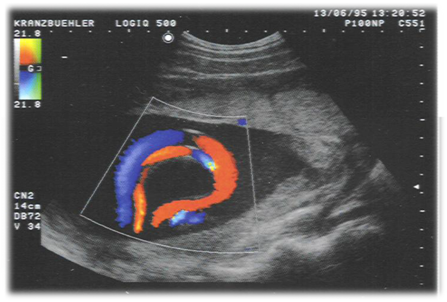
Since 1995 we have been engaged in dopplerography of arteries and veins and received patents of the technology for clinical interpretation of the detected pathology due to dopplerography.
In 2005 we developed a concept for arteriovenous balance, which radically changed views regarding measuring hemodynamic parameters.
For the last 10 years we have developed this concept in the applied technology, which enables to get more information from USD dopplerograms and interpret it for doctors in the comprehensive content.
Today parameters of the velocity and indexes are not sufficient and remain incomprehensible for practitioners.
Therefore, our technology provides creation of a set of additional measuring of amplitude, time duration and angels in dopplerograms for further processing.
Actually the work process of this technology can be divided into 4 stages:
- Obtaining and archiving dopplerograms of arteries and veins (a segment of 5 cardial cycles). 12 of 50 different vessels can be used in one case only from one research.
- Measuring of additional parameters in the automatic mode – amplitude, time and angular values in every chart of dopplerogram.
- Correlation mode of analysis of different arteries and veins and obtaining analytical indexes. Partly it can be performed in the automatic mode of post-processing after archiving dopplerograms.
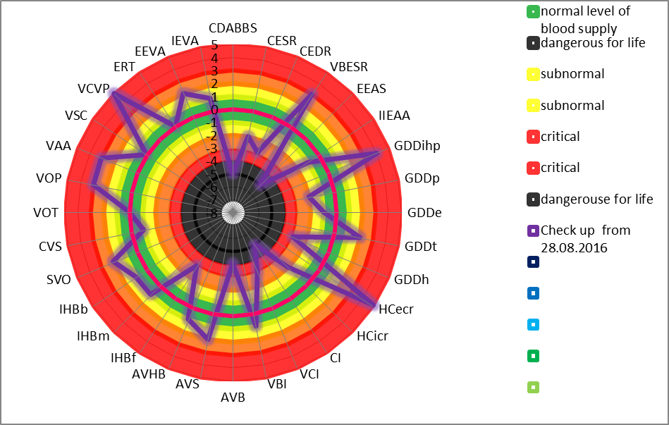
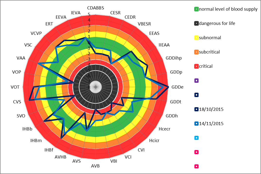
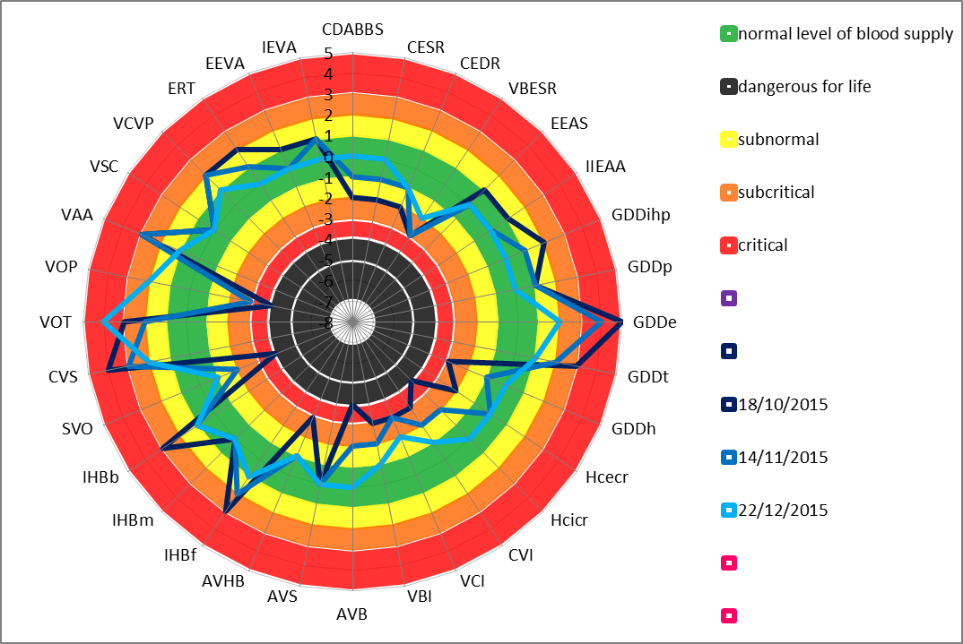
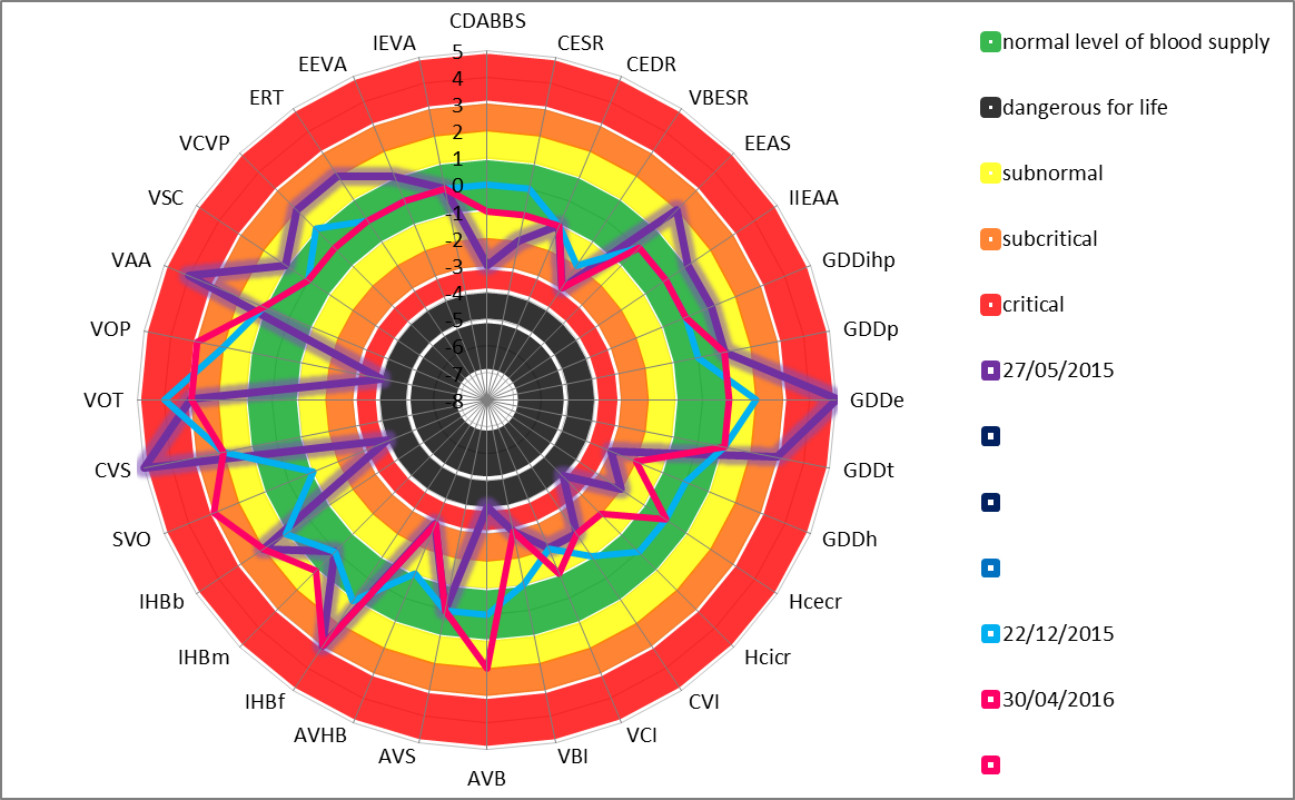
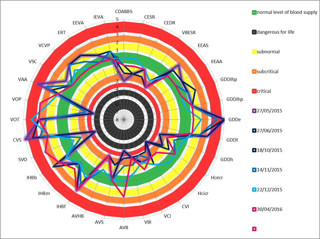
- Creation of the matrix for a circular histogram with the representation of accompanying text of conclusion with clinical interpretation.
Angiotherapy with the Angiomarker technology
The norm

The pathology
The dynamic of the angiotherapy

Ideology
Vascular USD 15 Years Ago: Stage of Observation and Learning
- Visualization of great vessels in vivo
- Admiring color mapping and duplex scanning
- Dopplerography of great and peripheral arteries with desire to understand the essence of USDG-indexes
- Transcranial scanning
- Gold standard is angiography
- Monitoring and detection of emboli – first stages – Pioner company
- Statistical way of static data accumulation
Vascular USD Today:
Visualization of Great and Peripheral Vessels’ Structure in vivo
- Practicing methodology for information gaining applying USD (3D), USDG, MRA, perfusion CT, and spiral CT (Radmir, Siemens, Materialize)
- 2D, 3D and 4D USD
- In-depth studying of USD structure and hemodynamic dysfunction of atherosclerotic plaques ( 2D, 3D)
- Early diagnosis of thrombosis
- Understanding the necessity of venous segment investigation
Noninvasive dynamic method (USAS is direct visual, USDG is indirect graphical).
The method is based on ultrasound radiation applying with using Doppler effect: the ultrasound wave changes its frequency on recoiling from movable blood elements, in particular erythrocytes.
The object of the investigation is a segment of the magistral artery or vein.
The level of the vascular system investigation is magistral vessels.
The method of results analyzing is quantitative-qualitative (digital and/or graphical).
US-angioscanning (USAS) is available in several modes of the ultrasound system work depending on its type and kind:
- modes of black and white imaging, the effect of color blood flow mapping (color angioscanning)
- and the energetic color blood volume coding, and the tissue perfusion colorization:
Using the modern US-system with the color coding and dopplerography, it is possible to receive more information about circulation state in magistral arteries and veins. In the mode of Doppler color mapping a qualitative evaluation of the lumina size, elastic-tonic and pulse features of the investigated segment of the artery, the vascular wall thickness, the organized nature of the blood flow with the diagnostics of disorganized areas in the form of turbulence and prognostication of the danger of the possible cerebral arteries embolia are carried out.

The modern US-system with the color coding of energetic Doppler effect offers the possibility of receiving the one-color picture of the circulation in organs, but it does not analyze the tissue type in organs, especially in zones of the intensive circulation, and does not differentiate the arterial and venous discirculation that is important for the individual pathogenic approach to the treatment policy.
The mode of energetic Doppler mapping allows to visualize cerebral arteries during the transcranial scanning and to evaluate the character of the arterial angioarchitectonics and the sinuosity of proximal segments.
Vascular USD Today:
Investigation of Great and Peripheral Vessels’ Function in vivo
- Linear velocity of artery and vein bloodflow
- Pulsatility (PI) and resistance (RI) indexes
- Turbulence indexes as embolia risk factors
- Creation of USDG – method based on technology of an open system with software polymorphism (Bioss and Spectromed companies)
- Renaissance of the idea of monitoring and detection of emboli applying conceptually new methodologies (Bioss company)
- Understanding the necessity of investigation of venous segment’s function
- Readiness to accept the idea of hemodynamic monitoring in critical conditions of the organism
Vascular USD Today:
Visualization of Microvasculature in Vivo
- Dopplerography of microvessels (Minimax)
- Laser dopplerography of microcirculation
- Digital optical capillaroscopy
- Understanding the necessity of examination of perivascular pressure, hydrodynamic conflicts, and arteriolar-venular balance
Vascular Diagnostics:
Questions for Tomorrow
- Necessity of developing methodology for dynamic investigations of vascular system
- Clinical analysis of results of different investigation methods for vascular system
- Change ascertaining dopplerography for dynamic one
We have started a stage of Applied Analytical Hemodynamics
- Analytical approaches to vascular system’s investigation
- New layer of modeling of hemodynamic changes in vascular systems in vivo under control of visualizing diagnostic methods
- In-depth studying of hydro- and hemodynamic laws, physics of ultrasound, and fundamentals of arterial and venous segments’ functioning for different types of angioarchitectonics
- Studying of hemodynamic reserve
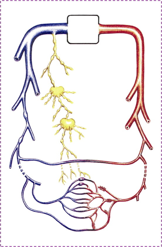 Ideology of Vascular Hemoduct
МАCROANGIOLOGY
- Investigation of pump function of the heart
- Investigation of great vessels of the head and extremities
- Investigation of veins of the head and extremities
- Investigation of appearance of blood filling and blood supply insufficiency on microcirculatory level as on the most distant segment of CVS (deep periphery, which is the most sensitive to ischemia)
- Decrease in pump heart function with evident reduction in blood filling in distal segments of arterial system and sludge phenomenon in microvasculature
- Impairment of elastic and tonic properties of great arteries’ vascular system resulting in formation of constrictive lesions of regional arteries
- Arteriovenous-hydrodynamic imbalance in case of apparent disorders in functioning of vascular system.
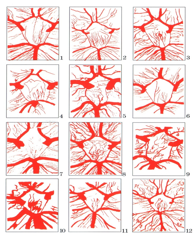
Medical Technology “Angioarchitectonics”
- Working out mathematical models of different-calibre architectonic type with calculations of hydraulic shock risk in case of atypical formation of arterial bed’s architectonics.
- Developing methodology for investigation and evaluation of architectonics requiring further works in the form of software for arterial U/S and MRI-technologists
- Developing algorithms of venous macroangioarchitectonics assessment.

МІCROANGIOLOGY
If signs of impairment in microcirculation picture are revealed, one should look for disturbance of minimum one of segments of the integrated closed CVS.
If microcirculation “picture” is healthy, one may speak of sufficiency and adequacy of CVS functioning in the “heart – great arteries – capillaries – great veins – heart” integrated segment
Investigation of microcirculation in finger and toe nail-bed is an arbiter of wellbeing of the whole vascular system
Modern Criteria of Vascular System Investigation
- Linear bloodflow velocity
- Vessel lumen and calibre
- Pressure (axial and matric)
- Tonus
- Elasticity of vessel wall
- Angioarchitectonics
- Arteriovenous balance